FREE SHIPPING ON ORDERS OVER $100
hello@theneighborhoodeyedoctor.com
What Are Retinal Vein Occlusions? Causes, Symptoms, and Treatment
Understand what retinal vein occlusions are, common risk factors, symptoms to watch for, how they are diagnosed, and available treatment options.
Dr.Ogechi Ukegbu
2/2/20262 min read
What Are Retinal Vein Occlusions? Causes, Symptoms, and Treatment
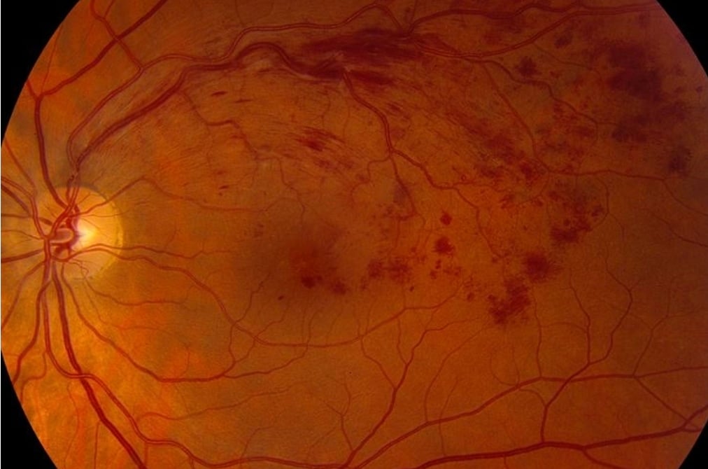
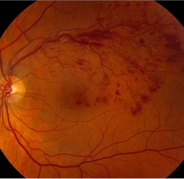
A retinal vein occlusion occurs when blood flow out of the retina is blocked, leading to swelling and vision changes.
Branch and Central Retinal Vein Occlusion (BRVO & CRVO)
Pathophysiology
Blood being pumped into the eye by the arteries must leave the eye, and the resultant high
blood pressure in the system ruptures the small capillaries connecting the arteries and
veins. Bleeding occurs in the retina, and the capillaries may be so damaged that they
become leaky, allowing serum to leak into the retina. The retina is normally like a dry
sponge of neurological tissue, and this leakage causes it to swell or thicken like a wet
sponge, preventing the retina from functioning correctly. When the retina, particularly the
macula (center of vision), is thickened from leaking fluid, this condition is called macular
edema. Further, the damage to the retina can be so severe that it dies from lack of oxygen, a
condition known as ischemia.
Complications of BRVO and CRVO
The complications of a Branch Retinal Vein Occlusion (BRVO) or a Central Retinal Vein
Occlusion (CRVO) are consistent with the damage inflicted on the eye. The most common
cause of decreased vision is leakage, resulting in macular edema. In many patients, this
condition resolves on its own over time. However, if it persists for three months or more,
laser or surgical therapy may be necessary. Another complication is the development of
new blood vessels. These vessels do not grow into the damaged retina where they are
needed, but rather into the vitreous cavity. Such vessels can bleed and partially or
completely fill the vitreous cavity, obstructing vision. Laser treatment or vitrectomy surgery
may be required, although in some cases, waiting and adjusting head positioning may help
restore vision. A particularly severe complication, especially in CRVO cases, is the
development of new vessels on the surface of the iris, known as rubeosis. These vessels can
obstruct the eye’s outflow channels (trabecular meshwork), leading to a difficult-to-control
form of glaucoma called neovascular glaucoma. Patients are closely monitored for the first
six months following a CRVO to detect this issue early.
Treatment and Clinical Studies
The Branch Vein Occlusion Study and the Central Vein Occlusion Study were conducted to
determine the effectiveness and timing of laser photocoagulation for treating macular
edema and neovascularization. The studies concluded that laser treatment was beneficial
for macular edema in BRVO cases but not in CRVO cases. Laser therapy was also found
effective for specific indications involving neovascularization of the retina and iris in both
diseases. Laser treatment presumably works by sealing leaking vessels and reducing oxygen
demand in damaged areas, thereby decreasing the stimulus for new vessel formation.
However, it does not reopen damaged vessels or restore normal blood flow to the retina.
Recent advancements include the use of anti-VEGF medications such as Avastin, Lucentis,
and Eylea, as well as long-acting intraocular steroids like Ozurdex, which have shown
promise in managing these conditions.
Disclaimer
The information contained in this document is for informational purposes only. Diagnosis
and therapy should be based on a thorough examination by and recommendations of a qualified eye care provider
Symptoms
Sudden blurred vision in one eye
Distorted or dark areas in vision
Causes and Risk Factors
High blood pressure
Diabetes
High cholesterol
Glaucoma
Diagnosis and When to See a Doctor
A comprehensive exam and imaging help identify the type and severity of occlusion.
Treatment and Management
Treatment may involve medication, injections, or referral to a retinal specialist.
Summary and Next Steps
Sudden vision changes should be evaluated promptly to protect vision.