FREE SHIPPING ON ORDERS OVER $100
hello@theneighborhoodeyedoctor.com
What Happens During a Retinal Detachment? Symptoms and Emergency Care
Learn the warning signs of retinal detachment, risk factors, why it is a medical emergency, and what to do if you experience sudden vision changes.
Dr.Ogechi Ukegbu
1/29/20262 min read
Retinal Detachment
Retinal detachment (RD) occurs when the retina, which is the neural tissue of the eye that
sees, separates from its nutritional supply of the retinal pigment epithelium (RPE) and
choroid. There are 3 different forms of retinal detachments: Rhegmatogenous Retinal
Detachment (most common form), Tractional Retinal Detachment, and Exudative Retinal
Detachment.
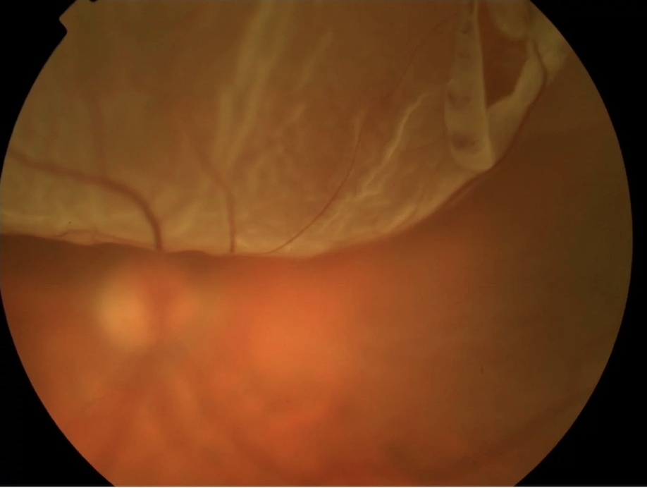
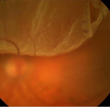
RETINAL DETACHMENT
The vitreous (jelly like substance in the eye) pulls on the retina, causing a retinal tear ("flap
tear"), and with constant contraction fluid enters the subretinal space and peels the retina
off causing a retinal detachment.
A rhegmatogenous retinal detachment which is the most common form and often referred
to just as a retinal detachment, arises from a retinal tear. Retinal tears develop either from
trauma, hereditary eye diseases, or from natural aging such as those that develop after a
posterior vitreous detachment. If not repaired, the retina is unable to function and begins
degenerating due to the lack of nutrition, leading to blindness. Surgery is necessary to
repair a rhegmatogenous detachment either by a scleral buckle, vitrectomy, or a
combination of both.
A tractional retinal detachment is seen often with proliferative diabetic retinopathy or with
proliferative vitreoretinopathy. It is the result of scar tissue that develops either from
abnormal blood vessel growth or from proliferating tissue that pulls on the surface of the
retina and causes the retina to separate from the underlying nutritive supply of the RPE and choroid
Function of the retina and blurred vision.
Surgery is necessary, often times by vitrectomy with a membrane peel, to remove the scar tissue that is tractionally causing the retina to detach.The last form is exudative retinal detachment. This is often times due to tumors or inflammatory disease processes resulting in fluid leakage arising from the RPE or choroid. The fluid leaks into the subretinal space and separates the retina from the RPE/choroid. Vision in this form of retinal detachment tends to be less impaired compared to the earlier two forms of retinal detachments due to fluid containing nutrients that are exuded from the RPE/choroid. The information contained in this document is for informational purposes only.
Diagnosis and therapy should be based on a thorough examination by and recommendations of a qualified eye care provider.